| Key
Facts |
- Rare non-langerhans
cell histiocytosis
- Pleural and perirenal
soft tissue thickening
- Diffuse septal
thickening
|
| Imaging
Findings |
Chest
radiograph
- Cardiomegaly, Kerley
B lines, blunting costophrenic angles (mimic CHF)
- Sclerotic bone
lesions
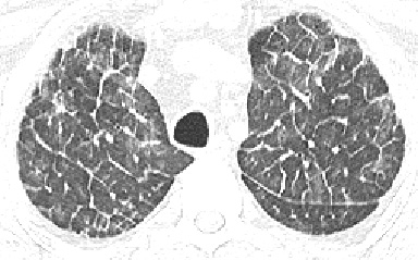
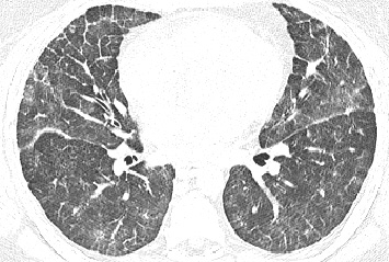
CT/HRCT
- Ground glass opacites
and centrilobular nodules
- Smooth septal thickening
- Diffuse pleural
thickening
- Encasement great
vessels or kidneys
|
| Differential
Diagnosis |
- CHF
- Lymphangitic tumor
- Diffuse pulmonary
lymphangiomatosis
- Pulmonary venoocclusive
disease
- Pulmonary alveolar
proteinosis
- Amyloidosis
|
| Pathological
Features |
- Proliferation of
non-langerhans cell histiocytes
|
| Clinical
Presentation |
- Middle age adults
- Pituitary involvment
presents with diabetes insipidus and exopthlmos
- Slow progression
of respiratory failure
- No known treatment
|
| References |
Wittenberg,
KH et al. Pulmonary involvement with Erdheim-Chester Disease: Radiographic
and CT Findings. AJR 174:1327-31, 2000
|