| Key
Facts |
- Upper lobe peripheral homogeneous consolidation
- During resolution, wavy lines paralleling chest wall
- Fever, weight loss, cough
- Rapid resolution
steroids
|
| Imaging
Findings |
Chest
Radiograph
- Homogeneous peripheral consolidation predominately
upper lung zones
- “Reverse butterfly pattern”
- “Photographic negative pulmonary edema”
- Normal heart size
- No pleural effusions or adenopathy
- May wax and wane like simple eosinophilic pneumonia
(Loeffler’s)
Resolution
- Inner edge of peripheral consolidation may form wavy
lines paralleling the chest wall
- Rapid resolution with steroids
- Little or no fibrosis with clearing
Recurrence
- Same place, same size, same shape
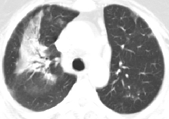
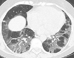
CT/HRCT
- Peripheral segmental subsegmental consolidation
- Contrast to chest radiography, lymph nodes may be enlarged
|
| Differential
Diagnosis |
- BOOP
- Sarcoid
- Churg-Strauss
- Mycoplasma
- Legionella
- Tuberculosis
- Parasites
- Drugs
- Penicillin
- Iodinated contrast
- Dilantin
- Methotrexate
- Ibuprofen
- Differentiation
- Tuberculosis
mimic: fever, weight loss, hemoptysis and upper lobe disease, tb often
cavitates
- Culture for parasites
- BOOP often lower
lobe predominant
- BOOP and sarcoid
may have peribronchial involvement
- Churg-Strauss:
one-third have pleural effusions, cardiac enlargement common either
dilatation or pericardial effusions, 70% have skin lesions,
|
| Pathological
Features |
- Alveoli flooded
with eosinophils and macrophages
- Bronchiolitis obliterans
in one-third
- Granulomas absent
|
| Clinical
Presentation |
- Cause unknown
- Typically middle aged women
- 50% have history asthma
- Cough
- Significant weight loss
- High fever
- Malaise
- SOB
- Occasional hemoptysis
- Eosinophilia in 90% (conversely
may be normal)
- PFT’s mild restriction
unless have asthma
Treatment
- Steroids
- Relapse common (50%)
with discontinuation steroids
|
| References |
Gaensler
EA , Carrington CB. Peripheral opacities in chronic eosinophilic pneumonia:
the photographic negative of pulmonary edema AJR Am J Roentgenol 128:1-13,
1977
Mayo JR, Muller NL, Road J, et al. Chronic eosinophilic pneumonia: CT
findings in six cases AJR Am J Roentgenol 153:727-730, 1989
Allen JN , Davis WB. Eosinophilic lung diseases Am J Respir Crit Care
Med 150:1423-1438, 1994
|