...about this most common x-ray examination.
Your Chest X-ray
 Roentgen discovered x-rays in 1895. This discovery would revolutionize the practice of medicine as it allowed, for the first time, to see inside the body without surgery. In recent years, new technology, computed tomography (CT or CAT) and magnetic resonance imaging (MRI) provide more and more details of the inner organs of the body. This allows earlier diagnosis of a wide variety of injuries and illnesses.
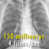
The chest x-ray is the most commonly performed radiographic exam. Approximately 45% of all radiographic exams are chest x-rays. Over 150,000,000 chest x-rays are done yearly in the U.S. at a cost of over 11 billion dollars.
Taking the X out of X-ray: 9 min movie demystifies x-rays and radiation. Narrated by famous inventor W.D. Coolidge of the General Electric Corporation. Dr. Coolidge was the inventor of the hot-cathode x-ray tube.
Inside Information: 8 min movie demonstrates how x-rays work and its use in police work, surgery, industrial engineering and automobile manufacturing.
Poor Quality
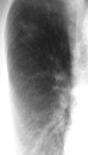 Unfortunately, although this is the most commonly performed examination, it often is inadequately performed. In an FDA survey, over 40% of the chest x-rays were judged inadequate by a panel of radiation physicists and physicians. Poor quality films markedly reduces the chance of finding abnormalities. Thus, it is important that the examination be a good one. A lung cancer was missed on this poor quality film. That's why quality is important!
Why Poor Quality
Chest x-rays are the most difficult of all radiographic examinations to perform because the chest contains tissues of such different consistency. The lungs are composed nearly entirely of air next to thick soft tissue and bone. Producing an image which adequately provides clear definition of all structures in the chest requires meticulous technique and attention to detail. The machine (film processor) used to develop the film must be working properly. This is rarely achieved. In the same FDA study, over 50% of the film processors surveyed were judged inadequate.
Good Quality
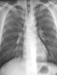 What is a good quality exam? First, ask if the facility is run by a board certified radiologist. The exam is more likely to be adequate because of their training. Ask what kind of equipment is used. Newer technology, such as computed radiography, or beam equalization radiography almost always ensures a good quality examination. Finally examine the film (see figure). Look behind the heart, the spine should be barely visible. The lungs should not be black. The blood vessels in the lung should be easily seen and crisp. Compare this good film with the poor quality film.
Radiation Dose
The dose of radiation from a chest x-ray is very small (0.25 mRad). Although this unit of measurement is probably unfamiliar, we all receive approximately 100 mRad (400 times that of a chest x-ray) yearly from cosmic rays and the trace radioactive minerals in rocks and building foundations. However, improperly used equipment can markedly increase the radiation dose. In an FDA survey, the range of x-ray exposures varied widely – one unit exposed patients to a whopping 120 mRad per x-ray!
Interpretation
 If possible, request that a board-certified radiologist interpret the films. There is no additional cost. A specialist in thoracic imaging is even better. While physicians other than radiologists also interpret films, they lack the training in radiation physics and radiation safety and the formal training in anatomy and physiology necessary for accurate film analysis. They also "look" at films incompletely. The schematic compares the eye movements of a trained radiologist versus an untrained physician. Note that the radiologist (on the left) has nearly completely examined all parts of the chest (in a few seconds) while the untrained physician (on the right) never looks at the outside portions of the chest. A small lung cancer will be easily missed by this physician. Multiple studies have shown that radiologists are far more accurate than untrained physicians in film interpretation. In a study by Pennsylvania Blue Shield, up to 40% of significant abnormalities on chest x-rays were misinterpreted.